

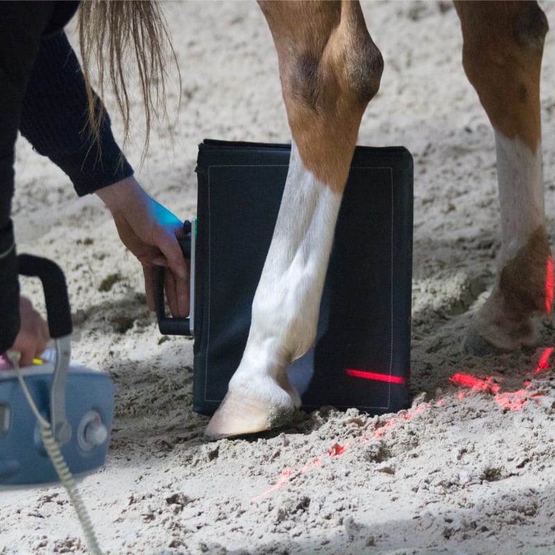
Portable X-Ray Technology For Horses
Conventional radiography (X-ray) is invaluable when it comes to diagnosing problems with the osseous structures of the horse. Vetwerx Equine offers portable digital radiography utilizing the most advanced technology. With our NEXT DR systems from Sound-Eklin, we are able to obtain precise clear pictures in seconds, enabling us to diagnose the most subtle of bone changes in the horse.
From acute emergency situations to scheduled pre-purchase exams, the portability of these machines allows us to provide top quality imaging at your location. In addition to the extremities, this equipment allows us to obtain clean images of the neck, head, shoulders, back, and stifles.
Digital X-Ray Case Studies
We take pride in educating our clients about all aspects of their horse's health. If you are interested in learning more about what our veterinarians can see with digital X-rays, take a look at the case studies below.
Pasture Injury
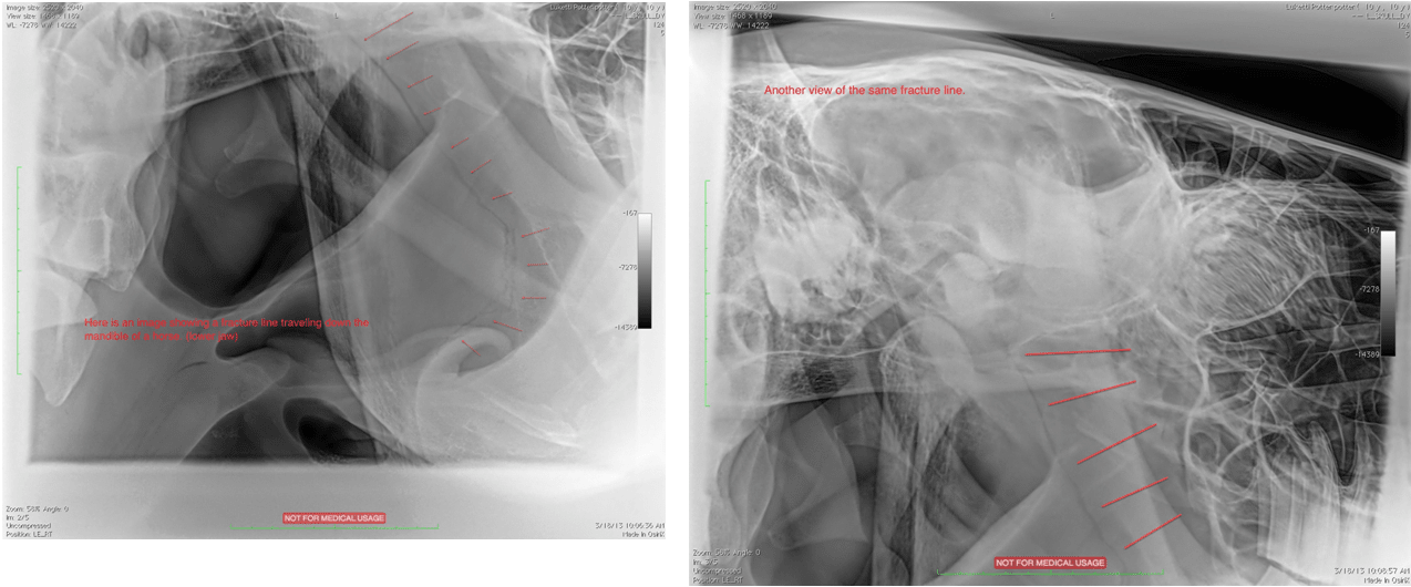
This is a horse that was observed falling while running in the pasture. He showed immediate signs of incoordination, difficulty eating, and neurological signs. He was treated with several days with anti-inflammatory drugs and radiographs were taken of the skull to determine if there were any fractures. A very large mandibular fracture was found that reached the temporomandibular joint.
Laminitis
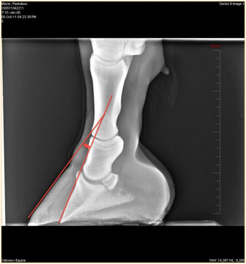
Here is a horse who has developed severe laminitis, also known as founder. The coffin bone has rotated away from the dorsal hoof wall, creating the angle that we've highlighted with the red lines. In a healthy horse, these lines should be parallel. In this case, the rotation is so severe that the coffin bone has gone through the bottom of the foot. The prognosis at this point is grave.
OCD (Osteochondritis dissecans)
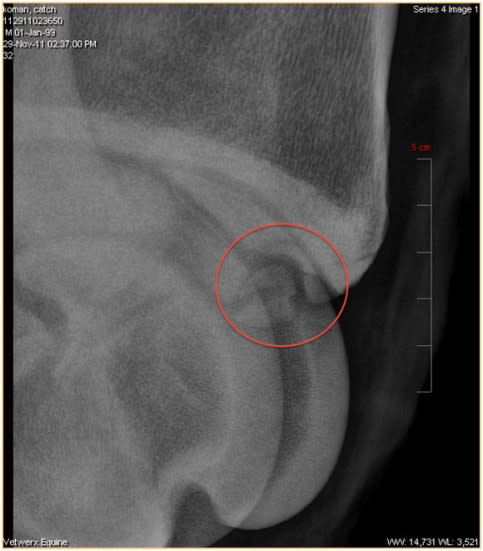
This is a horse that presented with a hind limb lameness and effusion of the tarsocrural joint in the hock. After localizing the lameness to the hock using joint flexions, radiographs were taken of the joint. You can see a fragment loose in the joint commonly known as OCD. This is an OCD lesion of the distal intermediate ridge of the tibia (DIRT).
Coffin Fractures in a 4-Month-Old Foal
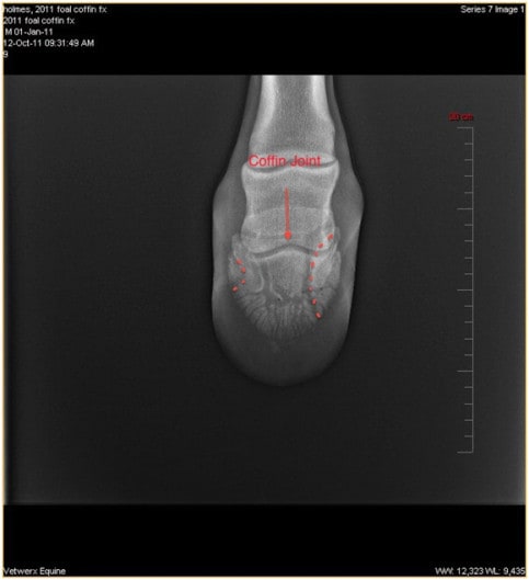
The smaller fracture to the left is known as a marginal fracture. The larger fracture on the right is a saggital fracture with communication to the coffin joint. Marginal fractures typically heal well with simple stall rest, however, communicating fractures require special care. An aluminum rim shoe was glued on, with stall rest for 3 months. Without this stabilization, the foal would invariably develop significant arthritis of the coffin joint, rendering the horse unusable for competition. With this course of treatment, the foal made a full recovery and will go on to compete.
Stifle Joint
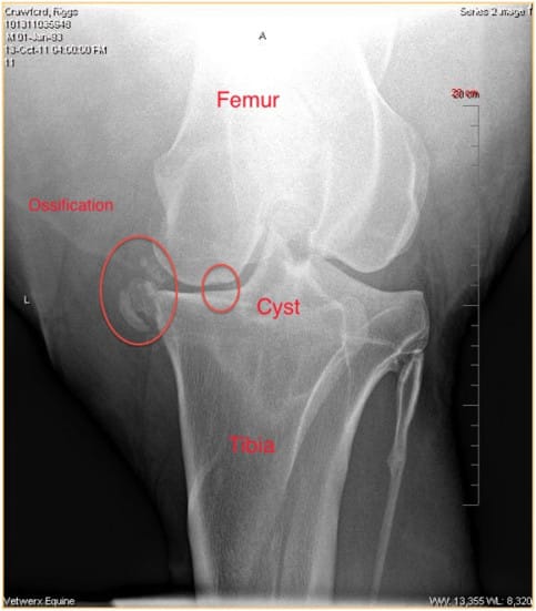
This is an older horse with chronic changes in the stifle joint. This horse had difficulty getting up and significant effusion of the stifle, particularly in the medial femorotibial compartment (MFC). Medial (inside of leg) is to the left. You can see ossification of the medial collateral ligament and meniscus, as well as a bone cyst in the MFC. The bone cyst is a form of OCD that can commonly be found here. These lesions typically appear at 1-2 years of age and routine screening of your young horses for these conditions can help start treatment early enough to prevent chronic arthritis like you see here.
Arthritis
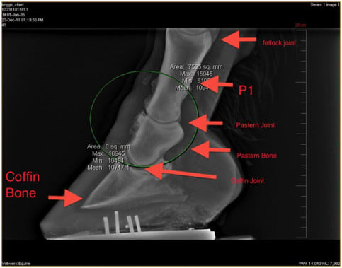
This is an older horse showing lameness in both front limbs. The horse was positive to flexion of the joints above. Diagnostic nerve blocks localized the lameness to below the fetlock joint. Radiographs revealed significant arthritis of the pastern joint, commonly known as high ring bone. You can see the bone proliferation surrounding the joint, having the appearance of "fluff". This is the bone remodeling as the arthritis progresses, causing inflammation of the joint, which is painful for the horse.